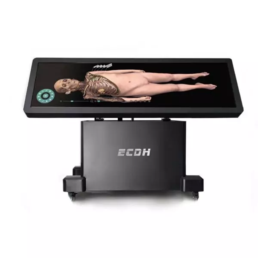
Precision and interaction are now the standard in anatomy education. DIGIHUMAN’s 3D virtual dissection table offers medical students and professionals a powerful platform that replicates human dissection in a digital, hands-on environment.
All-in-One Education System With Real Clinical Insight
What sets this system apart is its comprehensive database: over 3000 sectional images, 1800+ exercises, and a vast library of pathology and histology slides. Users can access actual clinical cases—more than 180 in total—complete with diagnostic images, case histories, and CT/MRI scans.
The 3D virtual dissection table enables users to manipulate anatomical layers, rotate perspectives, and simulate procedures with extreme precision. Whether it’s observing congenital abnormalities in the embryology module or zooming into a specific histological slice, the system supports in-depth, self-directed study.
DIGIHUMAN integrates touchpad and mouse functionality, making it easy for students to explore different magnifications and regions with just a few taps. This smooth user experience, combined with dynamic image control, allows learners to grasp medical concepts with greater clarity.
Enhancing Learning With Interactive Visualization
More than a display screen, DIGIHUMAN’s 3D virtual dissection table is a full-scale interactive learning environment. Students can wear 3D glasses for a stereo view of anatomical models, enabling better spatial awareness and retention. Educators benefit from customizable teaching flows and assessment tools that streamline course planning.
By bridging classroom instruction with clinical insight, DIGIHUMAN creates a seamless pipeline from theory to real-world practice.
Conclusion
DIGIHUMAN’s commitment to educational innovation shines through its 3D virtual dissection table. This tool empowers institutions with interactive content, real clinical cases, and unmatched detail—transforming how anatomy is taught and understood in modern medical education.